

How natural tanning pathways work: the biology explained
- 6 days ago
- 7 min read

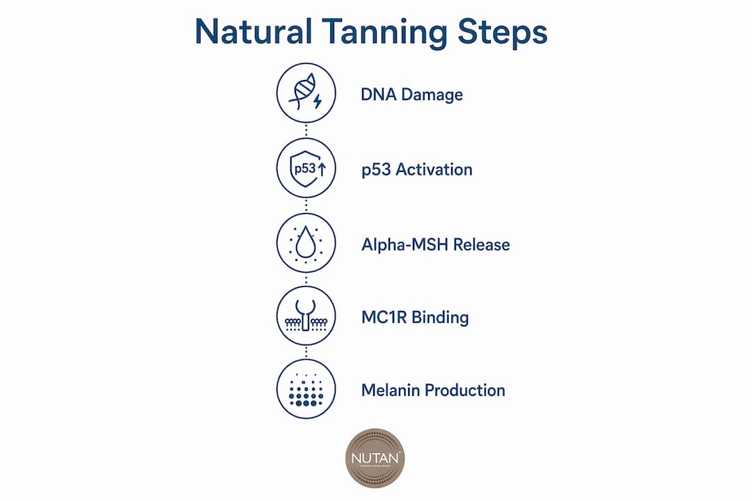
Your skin’s tanning response is a sophisticated biological defence system, not simply a cosmetic reaction to sunshine. When UV radiation reaches the skin, it triggers a precise chain of molecular events: DNA damage in keratinocytes activates the tumour suppressor protein p53, which drives expression of the POMC gene, producing alpha-melanocyte-stimulating hormone (alpha-MSH). That hormone binds to the Melanocortin 1 Receptor (MC1R) on melanocytes, setting off a signalling cascade that ultimately increases melanin production and redistributes pigment across the epidermis to shield your DNA from further harm.
The process unfolds in two distinct phases. An immediate response occurs within minutes of UV exposure, driven largely by oxidation and redistribution of existing melanin. Delayed tanning follows over days to weeks, fuelled by genuine new melanin synthesis. Both eumelanin (brown-black) and pheomelanin (yellow-red) are involved, though their ratio varies considerably between individuals, largely due to genetic differences in MC1R.
Table of Contents
How natural tanning pathways work at the molecular level
Understanding the tanning mechanism means following a relay of signals from the skin’s surface down to the pigment-producing machinery inside each melanocyte.
The initiating event: DNA damage and p53
UV-induced DNA damage in keratinocytes stabilises the p53 tumour suppressor protein within minutes of exposure. p53 then activates transcription of the proopiomelanocortin (POMC) gene. POMC is cleaved by proconvertase enzymes into several active peptides, most notably alpha-MSH, adrenocorticotropic hormone (ACTH), and the opioid beta-endorphin.
MC1R activation and the cAMP cascade
Alpha-MSH is secreted by keratinocytes and travels in a paracrine fashion to neighbouring melanocytes, where it binds the MC1R receptor. This binding raises intracellular cyclic AMP (cAMP) levels, activating the CREB/ATF1 transcription pathway and upregulating Microphthalmia-associated transcription factor (MITF), the master regulator of melanocyte identity and function.
MITF and tyrosinase: the pigment engine
MITF binds E-box sequences in the promoter regions of pigmentation genes, switching on tyrosinase, tyrosinase-related protein 1 (TYRP1), and dopachrome tautomerase. Tyrosinase is a copper-dependent enzyme that catalyses the rate-limiting conversion of tyrosine to L-DOPA and then to dopaquinone. From dopaquinone, the pathway branches: combination with cysteine produces pheomelanin precursors, while a separate route yields brown-black eumelanin.
Melanosome maturation and transfer
Melanin is synthesised inside melanosomes, which mature through four stages from a fibrillar matrix (Stage I) through progressive enzyme acquisition and pigment deposition (Stages II and III) to a fully melanised organelle (Stage IV). Mature melanosomes are then transported along microtubule networks using motor proteins, kinesin for outward delivery and dynein for return, towards the dendrites of the melanocyte. From there, they transfer to surrounding keratinocytes, where they form protective caps over the nuclei, physically shielding genomic DNA from further UV damage.
Timeline of the tanning response
Phase | Timing | Mechanism | Driver |
Immediate pigment darkening | Occurs soon after exposure | Oxidation and redistribution of existing melanin | UVA |
Persistent pigment darkening | Lasts for a limited duration | Dispersion of larger melanin granules | UVA |
Delayed tanning | Develops over a period of days to weeks | New melanin synthesis (melanogenesis) | UVB |
Melanin redistribution | Within one week | Upward migration of melanin to upper epidermal layers | UVB |
“The most significant change following UV exposure is a redistribution of melanin from the lower layer upwards to the middle layer of the skin, which is more dramatic in darker skin. This redistribution occurs across all racial groups examined and is critical to increased photoprotection.” — Journal of Investigative Dermatology, mechanisms of skin tanning study
Why tanning exists: protection, not pigmentation
Tanning is a biological defence response, not a cosmetic side effect of sun exposure. Melanin absorbs and scatters UV radiation, and its strategic placement as a cap over the keratinocyte nucleus means it intercepts incoming photons before they can damage the cell’s DNA. The photoprotective arrangement of melanosomes over nuclei is one of the most elegant adaptive mechanisms in human skin biology.
Melanogenesis is also genuinely costly. Tyrosinase is copper-dependent and tightly regulated, reflecting the metabolic investment required to produce melanin. The body does not run this process casually. Tight regulation of pigment production balances cellular protection against the energy expenditure involved, which is why tanning scales with UV dose rather than running continuously.
There is also a neuroendocrine dimension. POMC cleavage produces beta-endorphins alongside alpha-MSH. Beta-endorphins are opioid peptides that can influence mood and behaviour, which researchers have linked to UV-seeking tendencies in some individuals. The tanning pathway, in other words, is woven into a broader stress-response system that the skin shares with the central nervous system.
Pro Tip: Eumelanin provides substantially stronger photoprotection than pheomelanin. People with MC1R variants that favour pheomelanin production (often associated with red hair and fair skin) receive less DNA protection per unit of melanin produced, which partly explains their higher UV sensitivity.
Common misconceptions about the tanning process
Several persistent misunderstandings cloud how people think about tanning. Clearing them up makes the biology considerably easier to follow.
Biological tanning is not industrial tanning. Industrial leather tanning uses plant-derived polyphenols or chromium salts to chemically cross-link and harden collagen fibres in animal hides. Skin pigmentation involves no such chemistry. The shared word is a historical coincidence; the processes are entirely unrelated.
Immediate darkening is not new melanin. The colour change you see within minutes of UV exposure comes from oxidation of pre-existing melanin and redistribution of melanin granules, not from freshly synthesised pigment. True melanogenesis takes days.
UVA and UVB do different jobs. UVA drives immediate and persistent pigment darkening through oxidation. UVB is the primary driver of delayed tanning via the p53-POMC-alpha-MSH-MC1R pathway and genuine new melanin synthesis. UVA-induced colour tends to offer less photoprotective benefit than UVB-driven melanogenesis.
Eumelanin and pheomelanin are not interchangeable. Eumelanin is brown-black and strongly photoprotective. Pheomelanin is yellow-red and provides comparatively little UV defence. The ratio between them, determined largely by MC1R genetics, shapes both your visible tan and your actual level of sun protection.
Tanning involves both redistribution and synthesis. Within the first week after UV exposure, melanin redistribution from basal to upper epidermal layers accounts for most of the visible colour change. De novo synthesis becomes the dominant mechanism as basal melanin is depleted.
How NuTan® activates natural tanning pathways safely
The science of how natural tanning pathways work has opened a genuinely exciting avenue: triggering the same biological cascade without requiring high UV exposure. NuTan® transdermal tanning patches are built around this principle.
MC1R activation via MSH-ComplexB. NuTan® patches contain a proprietary Beta-melanocyte-stimulating hormone formulation, MSH-ComplexB, derived from a natural source. It binds and activates MC1R on melanocytes in the same way that alpha-MSH does naturally, initiating the cAMP-MITF-tyrosinase cascade and genuine melanin production.
Minimal UV requirement. Because the signalling pathway is already activated by the patch, only a small amount of sun exposure is needed to complete the tanning process. The result is a natural-looking tan that does not wash or rub off, because it is produced by your own melanocytes, not applied to the skin surface.
Downstream pathway engagement. By engaging MC1R directly, NuTan® activates the same downstream targets involved in pigmentation and cellular protection, including MITF upregulation and melanosome formation, that the body uses during natural UV-induced tanning.
Clinical relevance for MC1R variants. Research shows that MC1R variants reduce tanning efficacy in fair-skinned individuals. Approaches that activate the pathway at or downstream of MC1R offer a way to support pigmentation even where the natural UV response is limited.

NuTan® Triple Strength patches are available with worldwide delivery, offering a safe, needle-free way to work with your skin’s own biology for a natural, lasting glow.
Oxidative stress and antioxidant defences during tanning
UV exposure generates reactive oxygen species (ROS) as a direct by-product of photon absorption. These free radicals can damage lipids, proteins, and DNA independently of the direct UV-DNA interaction, adding a second layer of cellular stress during the tanning process.
Melanin itself acts as a free-radical scavenger, and its precursors and intermediates produced during melanogenesis also carry antioxidant activity. This dual role, as both a physical UV screen and a chemical ROS neutraliser, reinforces why the body invests energy in melanin production. Keratinocytes and melanocytes also upregulate enzymatic antioxidant defences, including superoxide dismutase and catalase, in response to UV-induced oxidative load. The balance between ROS generation and antioxidant capacity influences how well individual skin cells survive UV exposure and recover between sessions.
Why tanning responses vary so much between individuals
Genetic polymorphisms in MC1R are the single largest driver of individual variability in tanning response. MC1R is highly polymorphic, and loss-of-function variants are strongly associated with the red hair colour phenotype: fair skin, a tendency to freckle, poor tanning ability, and heightened UV sensitivity. People carrying these variants produce proportionally more pheomelanin and less eumelanin, reducing both visible tan depth and photoprotective benefit.
Beyond MC1R, body chemistry plays a broader role. Baseline melanocyte density, MITF expression levels, tyrosinase activity, and the efficiency of melanosome transfer to keratinocytes all vary between individuals and across different skin types. Research comparing tanning responses across ethnic groups confirms that while melanocyte density at the epidermal-dermal junction is remarkably similar across races, the distribution and redistribution of melanin differs considerably, with darker skin showing more dramatic upward melanin migration after UV exposure. Age, hormonal status, and certain medications can also modulate the pathway at multiple points.
Key takeaways
Natural tanning pathways are a UV-triggered biological defence system driven by p53 activation, alpha-MSH signalling through MC1R, and MITF-regulated melanin synthesis in melanosomes.
Point | Details |
p53 starts the cascade | UV-induced DNA damage in keratinocytes stabilises p53, which drives POMC transcription and alpha-MSH release. |
MC1R is the gateway | Alpha-MSH binding to MC1R raises cAMP, activating MITF and switching on tyrosinase for melanin synthesis. |
Early tanning is redistribution | Within one week of UV exposure, upward migration of existing melanin accounts for most visible colour change. |
Melanin caps protect DNA | Melanosomes position over keratinocyte nuclei to physically shield genomic DNA from further UV damage. |
MC1R variants affect outcomes | Genetic polymorphisms in MC1R shift melanin type towards pheomelanin, reducing tan depth and photoprotection. |
FAQ
What triggers the natural tanning response in skin?
UV radiation causes DNA damage in keratinocytes, which stabilises p53 and drives POMC gene expression. The resulting alpha-MSH binds MC1R on melanocytes, initiating melanin synthesis.
What is the difference between immediate and delayed tanning?
Immediate pigment darkening occurs within minutes via oxidation and redistribution of existing melanin, driven mainly by UVA. Delayed tanning develops over days through new melanin synthesis, primarily driven by UVB.
How do eumelanin and pheomelanin differ in terms of sun protection?
Eumelanin is brown-black and provides strong photoprotection, while pheomelanin is yellow-red and offers comparatively little UV defence. Your MC1R genetics largely determine which type your melanocytes produce.
Why do some people tan poorly despite sun exposure?
Loss-of-function variants in MC1R, common in fair-skinned and red-haired individuals, impair the signalling cascade needed for eumelanin production, resulting in limited tanning ability and greater UV sensitivity.
Can the tanning pathway be activated without high UV exposure?
Yes. Approaches that bind and activate MC1R directly, such as the MSH-ComplexB formulation in NuTan® patches, can initiate the same downstream melanin-producing cascade with only minimal sun exposure needed to complete the process.
Recommended